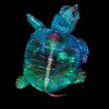


For forty-five years, the Nikon Small World Photomicrography Competition has celebrated the microscopic world and, in the process, allowed scientists and enthusiasts to show off the artistry of scientific imagery. Passionate micro-photographers from nearly 100 countries submitted over 2000 stunning images. Here, we present you some of the very best final selection with the winner photos of an embryo of turtle.
Teresa Kugler, the graduate who took this picture with her colleague Teresa Zgoda, says : “Microscopy lets us zoom in on the smallest organisms and building blocks that comprise our world–giving us a profound appreciation for the small things in life that far too often go unnoticed. It allows me to do science with a purpose.”
Via : My Modern Met

Fluorescent turtle embryo. Teresa Zgoda & Teresa Kugler (Campbell Hall, New York, USA). First Place. Stereomicroscopy, Fluorescence. 5x (Objective Lens Magnification).
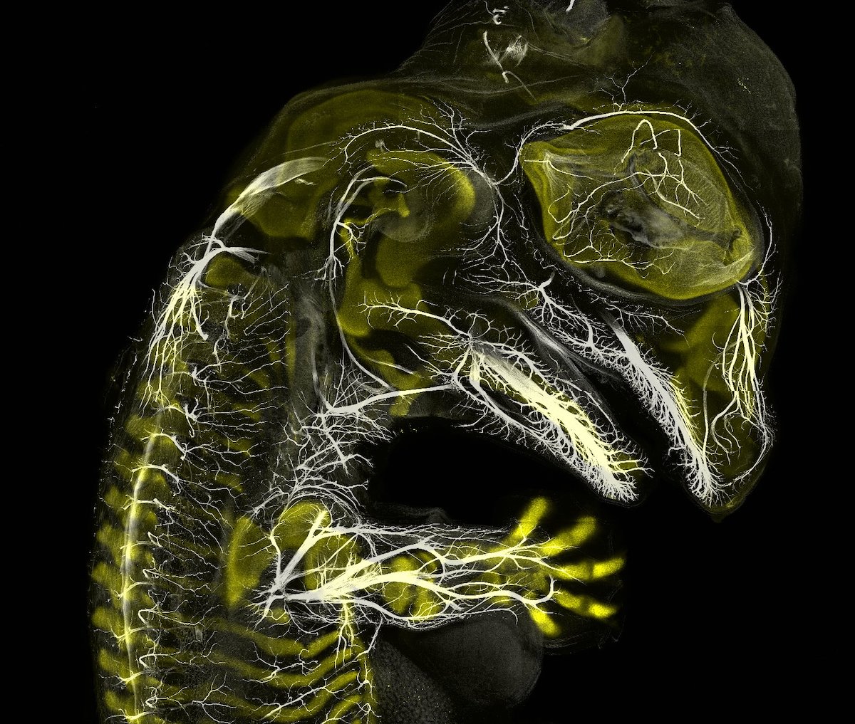
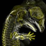
Alligator embryo developing nerves and skeleton. Daniel Smith Paredes & Dr. Bhart-Anjan S. Bhullar (Yale University, Department of Geology and Geophysics, New Haven, Connecticut, USA). Third Place. Immunofluorescence. 10x (Objective Lens Magnification).
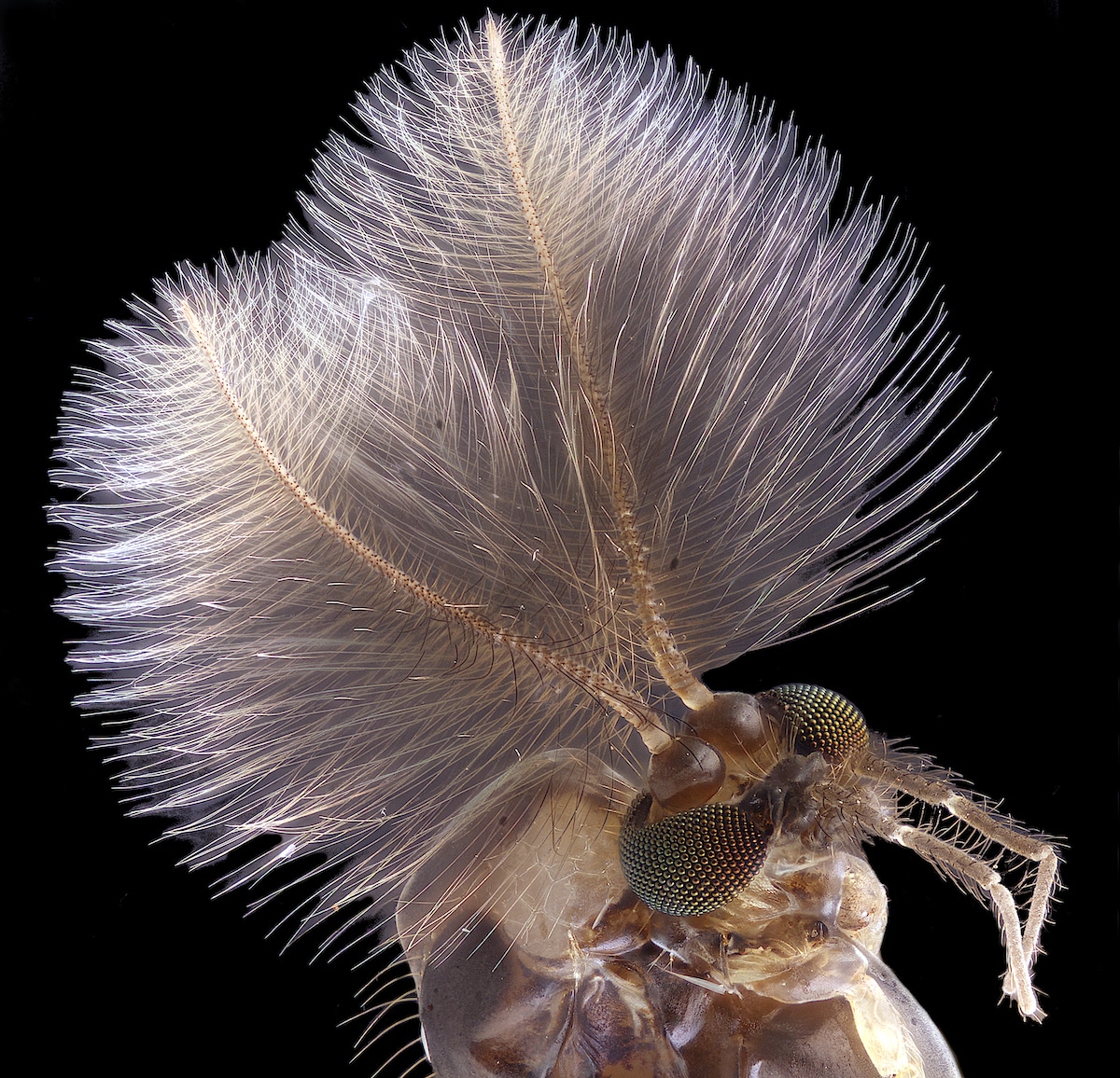
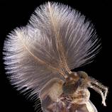
Male mosquito. Jan Rosenboom (Universität Rostock, Rostock, Mecklenburg Vorpommern, Germany). Fourth Place. Focus Stacking. 6.3x (Objective Lens Magnification).
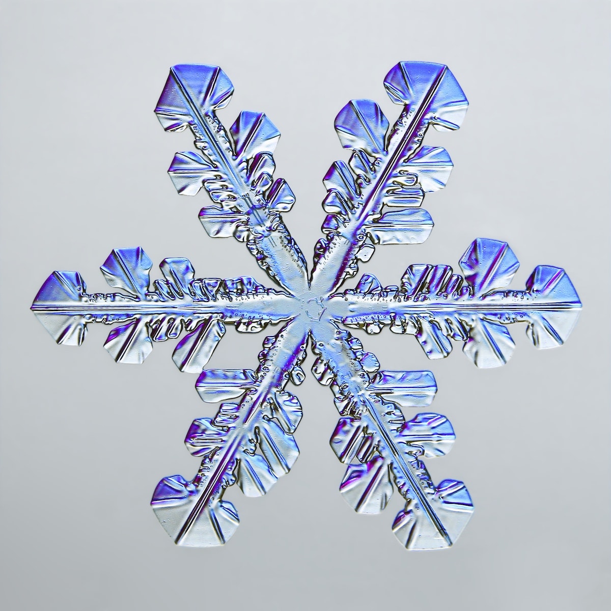
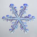
Snowflake. Caleb Foster (Caleb Foster Photography, Jericho, Vermont, USA). Fifth Place. Transmitted Light. 4x (Objective Lens Magnification).
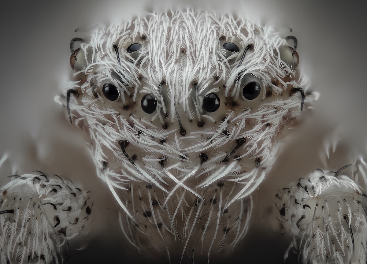
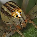
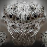
Small white hair spider. Javier Rupérez (Almáchar, Málaga, Spain). Sixth Place. Reflected Light, Image Stacking. 20x (Objective Lens Magnification).
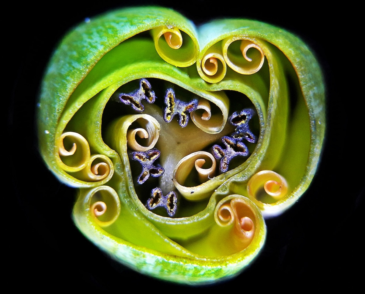
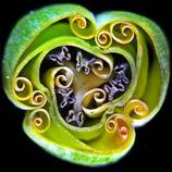
Tulip bud cross section. Andrei Savitsky (Cherkassy, Ukraine). Ninth Place. Reflected Light. 1x (Objective Lens Magnification).
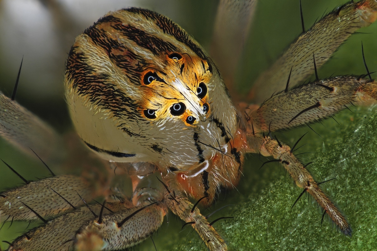
Female Oxyopes dumonti (lynx) spider. Antoine Franck (CIRAD – Agricultural Research for Development, Saint Pierre, Réunion). 14th Place. Focus Stacking. 1x (Objective Lens Magnification).
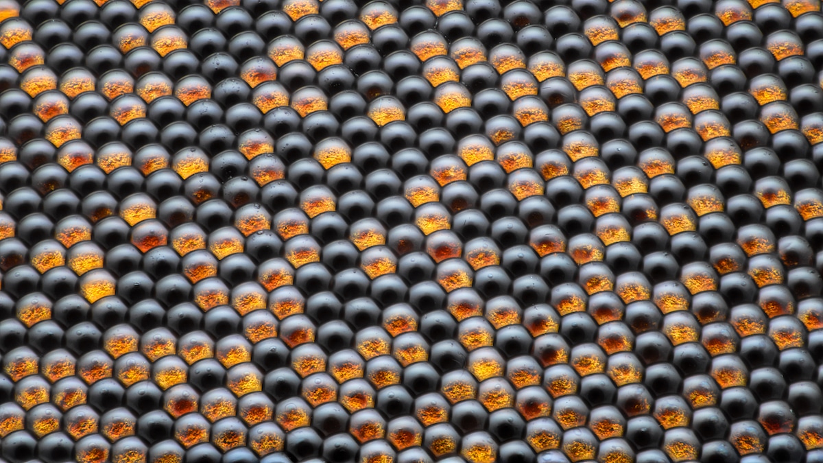
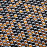
Housefly compound eye pattern. Dr. Razvan Cornel Constantin (Bucharest, Romania). 16th Place. Focus Stacking, Reflected Light. 50x (Objective Lens Magnification).
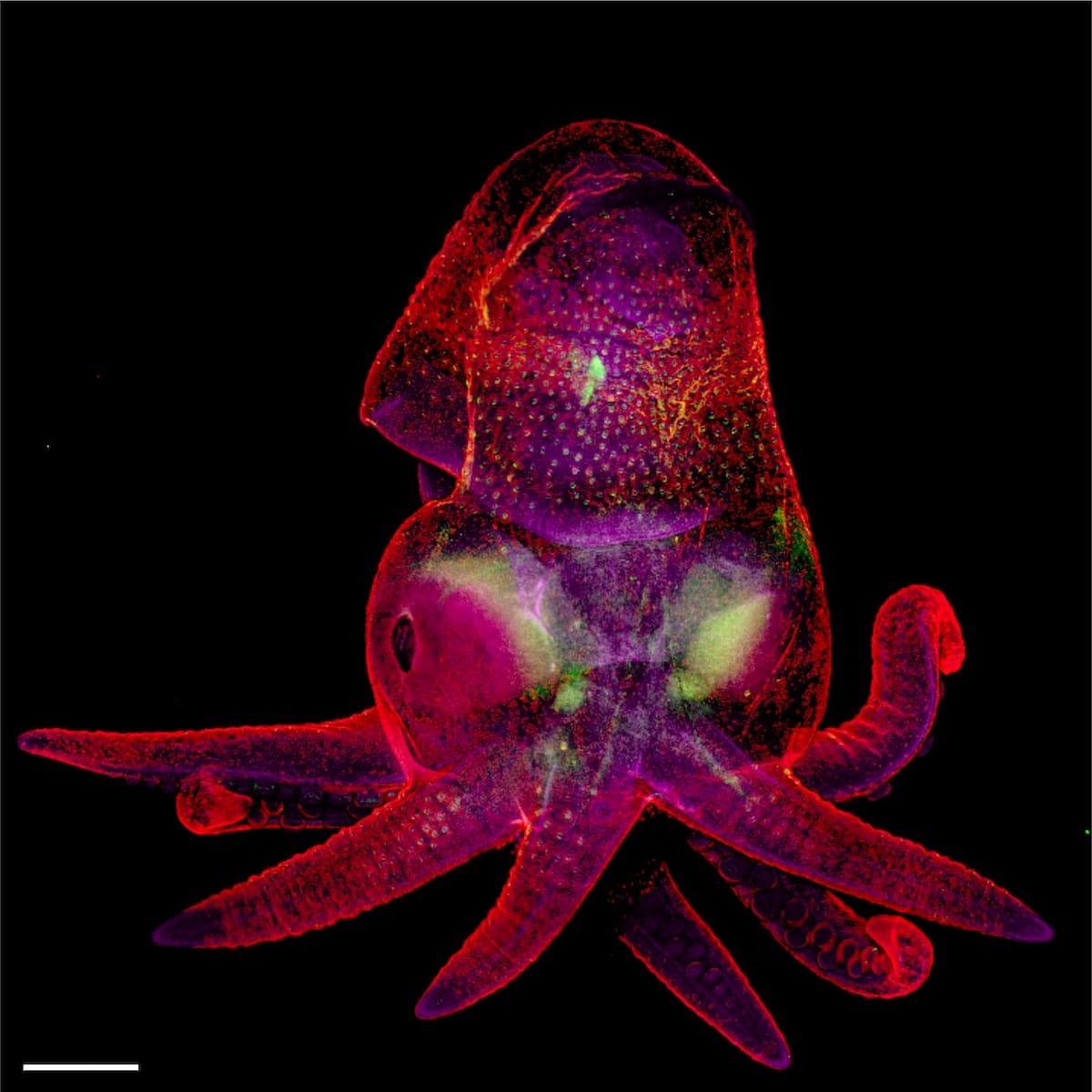
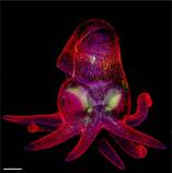
Octopus bimaculoides embryo. Martyna Lukoseviciute & Dr. Carrie Albertin (University of Oxford, Weatherall Institute of Molecular Medicine, Oxford, Oxfordshire, United Kingdom). 19th Place. Confocal, Image Stitching. 5x (Objective Lens Magnification).